

When it comes to talking about causes and treatment of Laminitis, there are many opinions “out” there, and sometimes it appears they contradict each other. How can this be? Does that mean only some views are correct, and consequently the others must be incorrect?
Based on my experience over the last two decades, I have concluded that the confusion and different opinions we read are the result of mixing up different event mechanisms that lead to a situation we call laminitis. In other words, we are not talking about the same “thing”.
The common denominator is the word Laminitis and on the first glance they look similar on x-rays. While acute, the horse presents the same way, aka fronts camped out, hinds standing under, high pulse, stiff gait, etc. The management of the acute phase (< 72h) would typically be the same, too.
However, how the horse got to this stage is different. As far as I am concerned, there are two fundamentally different types of scenarios, 1) chemical overload of some sort, and 2) mechanical overload.
In the true sense of the first scenario, the hoof is healthy prior to the event. That means it is without wall separation and the consequences of the trigger event allows the hoof-capsule to potentially separate.
In the second scenario, the separation develops first as a result of mechanical deformation and overload, while the inflammation part is triggered later – often years apart.
As chemical overload I regard scenarios like insulin overload, carbohydrate overload, toxicity response from retained placenta, etc. They all create a toxicity effect that tends to affect the whole horse. In the hoof it inflames the laminae, and the laminar dermis and epidermis appear to separate. Interestingly enough, this tends to happen without physical damage of the lamellar structure, aka no ripping and breaking. Theoretically, this should happen within a healthy and tightly connected hoof-capsule (HC), free of distortion, because we would like to assume that the hoof was healthy prior to such event. In the first occurrence of such event within a healthy HC, there simply is no space for P3 to to go anywhere, due to the close proximity of inner hoofwall and P3. Unfortunately, we rarely find those healthy hooves to begin with.
The insulin and carbohydrate overload scenarios are have been studied and created in a pseudo-in-vivo environment, where a horse is forced to receive large amounts of these substances within a very short period of time, that in real life with normal boarding conditions would be extremely hard, if not impossible, to achieve in a 24h period.
Due to the “letting go” of the laminar connection, if one was to dissect hooves from this scenario, one could expect that the HC pulls away from the laminar corium relatively easily.
The mechanical overload is by far more common in real life. Typically this starts in the back half of the hoof where discomfort is allowed to develop in the hoof. This can start as early as a few months after birth. The horse shifts weight away from that area and transfers it towards the front half of the hoof – 24/7. This paves the way for chronic overstressing and eventually stretching of the laminar connection, and sometimes even the laminar corium. Those horses commonly present symptoms like fast growing heels, steep pastern alignment, short stride, toe-first landing, possibly stumbles easily, tight muscles around the shoulders. Eventually we see flare in the toes and radiographically we would see P3 and the inner hoofwall are no longer parallel. That means the laminar connection is getting widened. All the while the horse does not show any signs of acute laminitis, but it would most likely be diagnosed with “signs of chronic laminitis”.
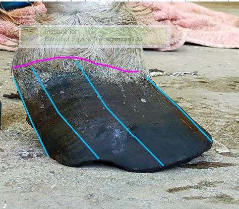
The space between P3 and the hoof-capsule is filled out with more laminar horn, often referred to as “laminar wedge”. More laminar horn over a wider space does not make the hoof stronger, but rather the opposite occurs. Usually the harmonic laminar arrangement is disturbed, making the lamellae look rugged and “ripped”. Nevertheless, when dissecting such a hoof, it is typically very hard to separate the laminar horn from its corium – often the corium rips off the bone instead.
Typically, a trigger event of some sort (trauma, dietary disturbance, change of ground conditions, change of hoof hydration) occurs, and promotes and acute laminitis. Many horses only get x-rayed at this point, which then shows separation on a hoof with acute laminitis. A very common reason, why it is believed that the laminitis caused the separation.
The problem now is that we are trying to explain, and treat, this second scenario with the reasoning from the first scenario. However, it is to note that a hoof with mechanical distortion and toe overload will have a much lower trigger threshold to chemical overload. The long-term solution then is to lift that trigger threshold and focus on reducing the HC distortion.
